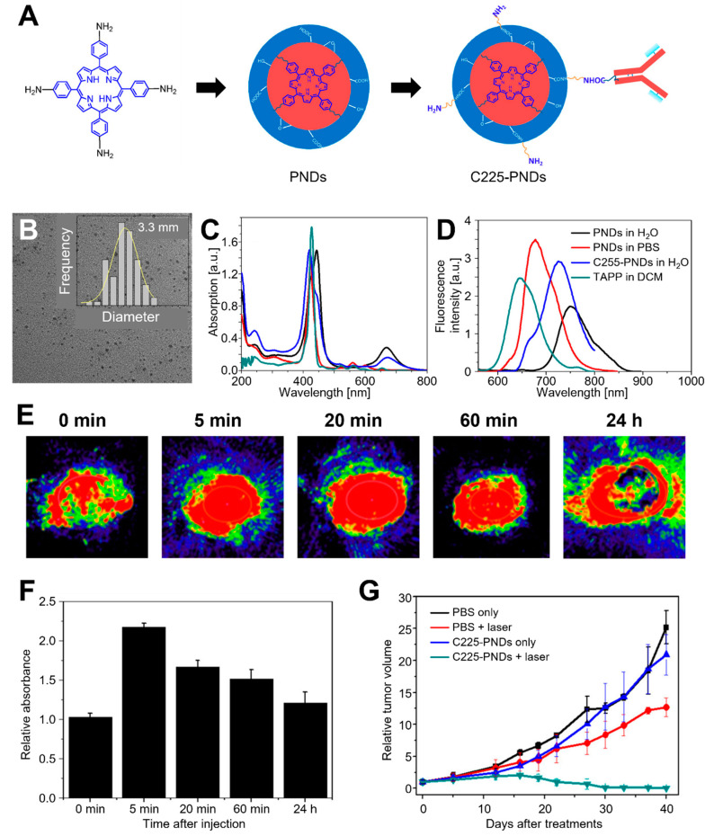
Figure 8.
C225–PNDs for PAI and in vivo breast cancer ablation. (A) Proposed formation pathway of PNDs and the synthetic routes of C225–PNDs. (B) TEM image of PNDs with a corresponding size distribution histogram. (C) UV–vis spectra and (D) emission spectra (λex = 440 nm) of PNDs, C225–PNDs, and 5,10,15,20-tetrakis (4-aminophenyl) porphyrin (TAPP). (E) PA imaging and (F) relative PA intensity of C225–PNDs in the tumor at different time points. (G) Relative tumor volume. The images are reproduced with permission from ref. [165].

