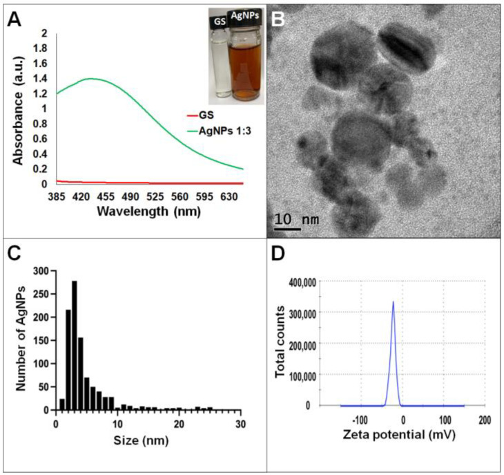
Figure 3.
Silver nanoparticles obtained with the optimized protocol using the supernatant of G. sessile. (A) UV-Vis analysis of AgNPs-GS after 72 h of reaction, (B) TEM micrograph showing morphology of NPs, (C) size distribution histogram of AgNPs, (D) Zeta potential. Inset in (A) shows the fungal supernatant and synthesized NPs. GS = Ganoderma supernatant.

