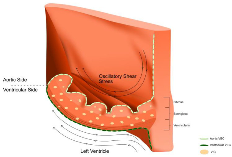
Figure 3.
An aortic valve leaflet comprises an extracellular matrix that is organised into three layers; the ventricularis is on the ventricular side, the fibrosa is on the aortic side and the spongiosa is sandwiched in between these two layers. Valvular endothelial cells (VECs) line the ventricularis and fibrosa. Valvular interstitial cells (VICs) are found in all the layers. The aortic valve is exposed to a complex and harsh environment; the ventricularis side is exposed to laminar flow and shear stress, whereas the fibrosa side is exposed to oscillatory flow patterns and lower shear stresses.

