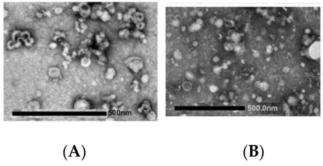
Figure 8.
Transmission electron micrographs of exosome samples collected from cell culture and visualized after negative staining. In (A) the preparation is a population of large and small vesicles; the morphology is not uniform, some vesicles appear with a cup-like structure some with a deformed shape. Slight clumping can also be observed. In (B) vesicles are surrounded by microparticles, precipitates, and impurities generated in the stain or during preparation.

