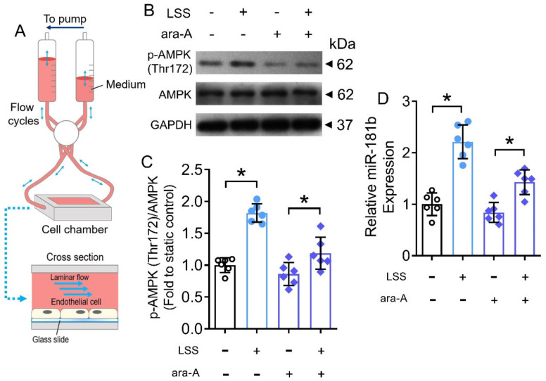
Figure 7.
Effects of LSS on AMPK/miR-181b axis in endothelial cells. (A) Schematic diagram on the design of ibidi flow system and cross section of flow chamber. (B) Representative Western blots, and (C) quantification of Western blotting on expression of AMPK and p-AMPK at Thr172 in HUVECs exposed to LSS for 12 h. n = 6 per group. (D) RT-PCR on miR-181b expression in LSS-exposed HUVECs. n = 6 per group. Data are mean ± SD. * p < 0.05 (Brown-Forsythe and Welch ANOVA and Games-Howell’s multiple comparisons).

