Figure 3.
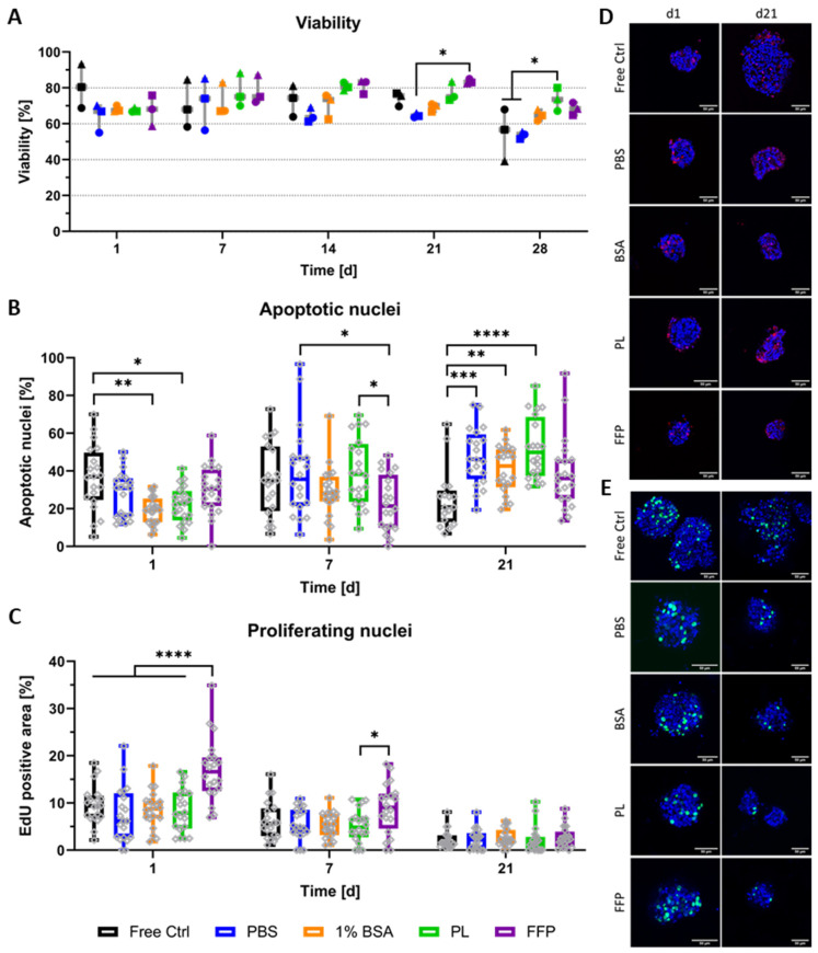
Viability and proliferation of bioprinted NICC in supplemented algMC. Analysed groups are free control NICC in suspension culture and NICC bioprinted in PBS-algMC, BSA-algMC, PL-algMC, and FFP-algMC. (A) Semi-quantitative analysis of live/dead staining, the different symbols represent the different experiments depicted, mean ± SD, n = 3, >25 NICC each; (B) Quantitative analysis of percent apoptotic nuclei per islet, mean ± SD, n = 1, 20 NICC; (C) Quantitative analysis of proliferating nuclei. Mean area of proliferating nuclei in percent of total islet area, mean ± SD, n = 1, 20 NICC; (D) TUNEL staining for apoptotic nuclei in bioprinted NICC, representative images of samples fixed on d1 and d21 after bioprinting, scale bars = 50 µm; (E) EdU staining for nuclei of cells that proliferated within 24 h before fixation, representative images of samples fixed on d1 and d21 after bioprinting, scale bars = 50 µm. Grey dots in a-c indicate individual islets, significances indicate * p < 0.05, ** p < 0.01, *** p < 0.001, **** p < 0.0001.

