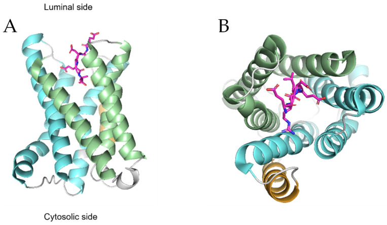
Figure 3.
Structural organization of KDELR. The images show the side (A) and top view (B) of Gallus gallus KDELR2 according to the PDB database coordinates (PDB DOI: 10.2210/pdb6ZXR/pdb 27 April 2022). The two helical bundles made up of the first three and last three helices are shown in light green and light blue, respectively. The fourth transmembrane helix, which connects the two bundles, is shown in light brown. The RDEL ligand, represented in sticks, is located in the pocket formed by the TM1-3 and TM5-7 helices.

