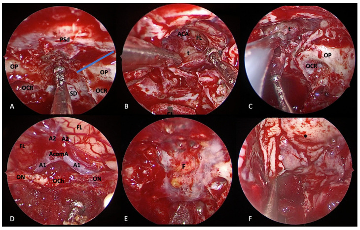
Figure 7.
Extended endoscopic endonasal transplanum/transtuberculum approach for removal of tuberculum sellae meningioma. (A) Margins of bone removal: laterally, the optic nerves and the optocarotid recess; inferiorly, the sellar floor; anteriorly, the bone removal is tailored on tumor extension. Extracapsular dissection (B) and internal debulking (C) of the tumor. Surgical field view after tumor removal (D). Closure is performed using fat (E), nasoseptal flap (F), and fibrin glue. Tumor (t); dura of the planum sphenoidale (PSd); optic protuberance (OP); optocarotid recess (OCR), sellar dura (SD); frontal lobe (FL); anterior cerebral artery (ACA); optic nerve (ON); optic chiasm (OCh); fat (F); nasoseptal flap (*).

