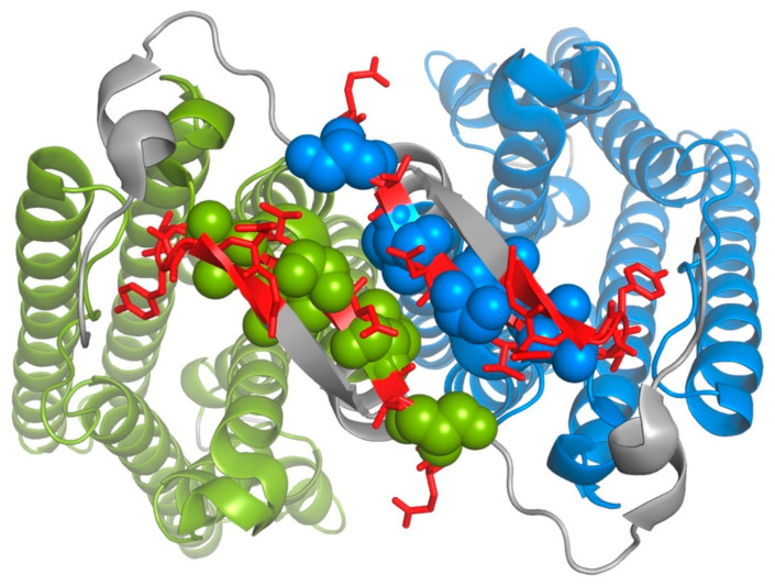
Figure 3.
Accessibility of the N-terminal region of Ste2p mapped onto the structure of the ligand-free Ste2p dimer (PDB code: 7QB9) [37]. Sites in the N-terminal region where introduced cysteine substitutions could be labeled by MTSEA-biotin are shown as red sticks. Sites in the N terminus that could not be labeled by MTSEA-biotin are shown as green and blue spheres for the two different monomers. Sites in the N-terminal region that were not tested for MTSEA-biotin labeling are shown in grey. Portions of Ste2p other than the N terminus are depicted using cartoon representation, colored green and blue, respectively, for the two monomers.

