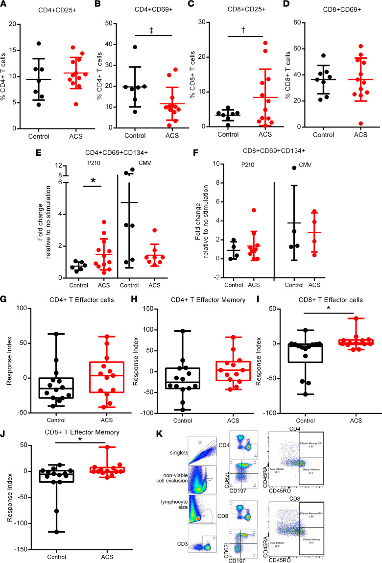
Figure 1. Intrinsic T cell response to P210 peptide in human PBMCs.
Human PBMCs from control patients and acute coronary syndrome (ACS) patients were cultured for 16 hours for the AIM assay with no stimulation or stimulated with P210 peptide or CMV pooled peptides. (A–D) Activation state of PBMCs without peptide stimulation. (E and F) AIM+ cells in response to P210 or CMV peptide pool. (G–J) PBMCs were stimulated with P210 peptide for 72 hours, and cells were stained for T effector and memory markers. The box plots depict the minimum and maximum values (whiskers), the upper and lower quartiles, and the median. The length of the box represents the interquartile range. (K) Gating scheme for T effector and memory cell analysis. Mann-Whitney U test except for C and E, 2-tailed t test. ‡P = 0.07; †P = 0.05; *P < 0.05. (A–F) Control n = 7–8, ACS n = 12; some samples/treatments did not have detectable AIM+ cells so ratio could not be determined. (G–J) Control n = 14, ACS n = 13.

