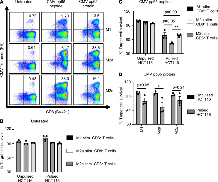
Figure 6. CD206+ macrophages can efficiently cross-present soluble CMV pp65 Ag.
(A) Dot plots indicate expression of CMV-specific TCR on CD8+ T cells as analyzed by MHC I tetramer staining. CD8+ T cells were stimulated with untreated MDM or MDM pretreated with CMV pp65 short peptide, or they were stimulated with MDM pretreated with CMV pp65 protein. Numbers indicate percentage of CD8+CMV+ T cells. Data are representative of 3 donors. (B–D) Data show cytotoxic activity of differentially stimulated CD8+ T cells, wherein autologous CD8+ T cells were cocultured with untreated (B), CMV pp65 peptide pretreated (C), or CMV pp65 protein pretreated (D) MDM subsets. Bar diagrams show percentage survival of target HCT116 cells, cocultured with differentially stimulated CD8+CMV+ T cells, normalized to HCT116 cells alone. HCT116 cells were either unpulsed or pulsed with CMV pp65 peptide before coculturing with stimulated CD8+ T cells. (B–D) Data show mean ± SEM (n = 3 donors). Unpaired Student’s t test was used. For multiple comparisons, Holm-Šídák test was performed (C). *P < 0.05, **P < 0.01.

