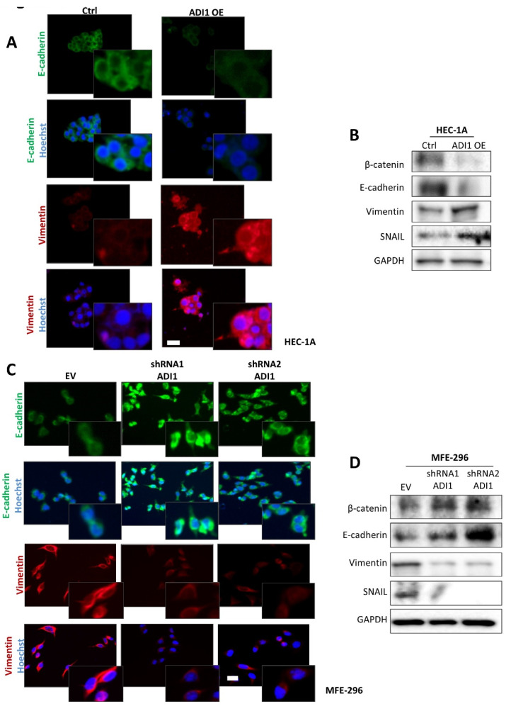
Figure 4.
Ectopic modification of ADI1 levels modulates epithelial features. (A) Representative images of immunofluorescence against E-cadherin and vimentin in HEC-1A cells transfected with ADI1 over-expression vector. Magnification images of framed regions of the samples are shown. Scale bars: 20 μm. (B) Representative images of western blot analysis of E-cadherin, β-catenin, vimentin and SNAIL in HEC-1A cell line transfected with ADI1 over-expression vector. GAPDH was used as a loading control. (C) Representative images of immunofluorescence against E-cadherin and vimentin in MFE-296 cells infected with lentiviral shRNA1 ADI1 and shRNA2 ADI1 plasmids. Magnification images of framed regions of the samples are shown. Scale bars: 20 μm. (D) Representative images of western blot analysis of E-cadherin, β-catenin, vimentin and SNAIL in MFE-296 cells infected with lentiviral shRNA1 ADI1 and shRNA2 ADI1 plasmids. GAPDH was used as a loading control. The whole western blots were shown in File S2.

