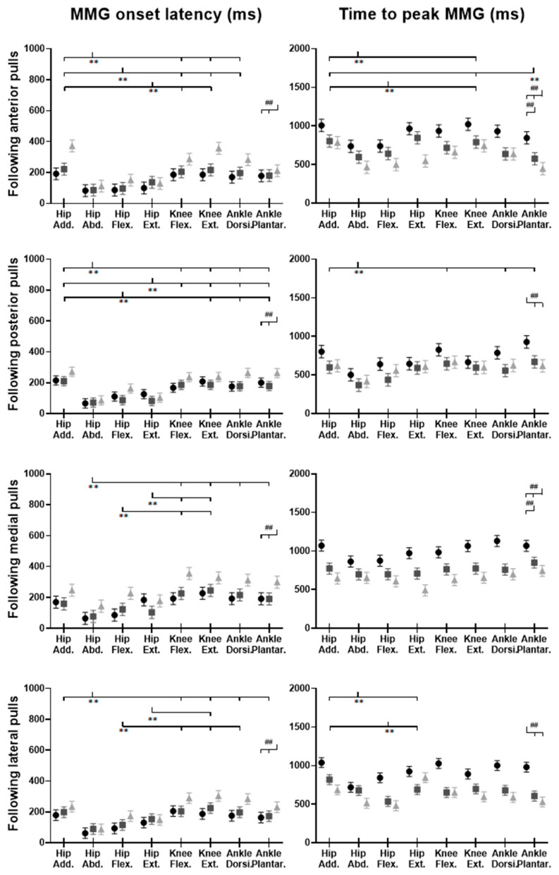
Figure 15.
The MMG onset latencies and time to peak MMG amplitude for eight dominant-leg muscles following unexpected horizontal perturbations (mean ± SE, n = 12). (Note: Hip Add.: adductor magus; Hip Abd.: gluteus medius; Hip Flex.: iliopsoas; Hip Ext.: gluteus maximus; Knee Flex.: semitendinosus; Knee Ext.: rectus femoris; Ankle Dorsi.: tibialis anterior; Ankle Plantar.: gastrocnemius medialis. MMG: mechanomyography; SE: standard error;  or
or  : pairwise comparison. Significant differences in post hoc pairwise comparisons (p < 0.05) were indicated by the: ** for the main effect of muscle factor; ## for the main effect of magnitude factor).

