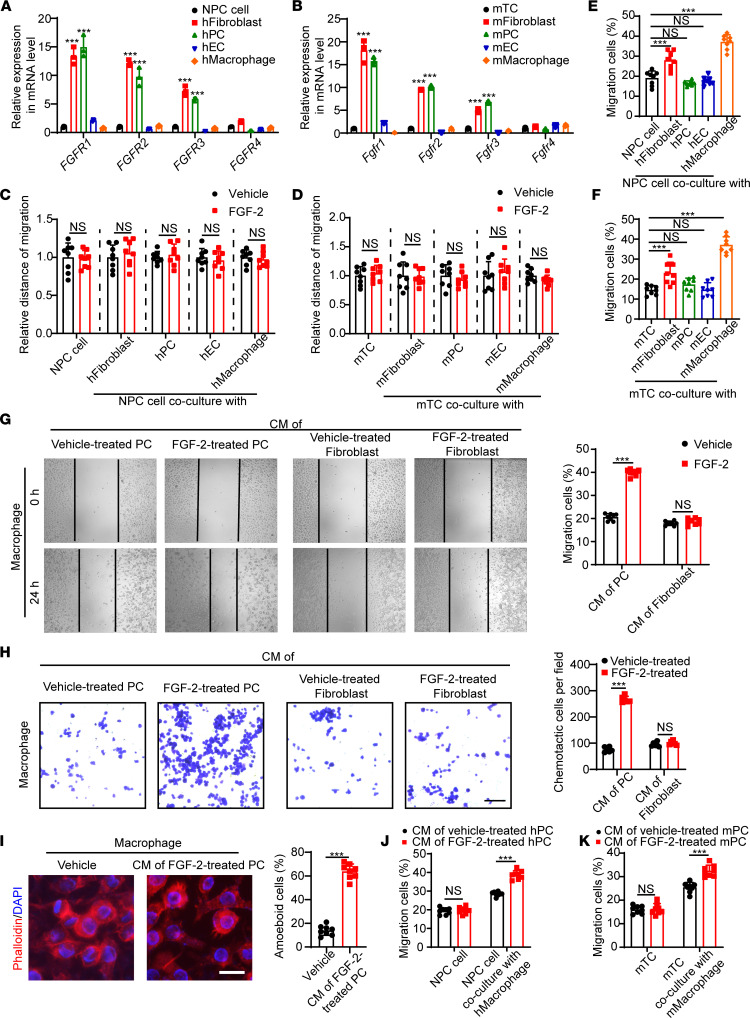
Figure 3. Pericyte-dependent mechanism of FGF-2–induced macrophage activation.
(A and B) qPCR quantification of human and mouse FGFR1, FGFR2, FGFR3, and FGFR4 mRNA levels in various cell types, including 5-8F NPC cell line, hTERT-immortalized dermal fibroblasts, isolated primary pericytes, HUVEC endothelial cells, THP-1 monocyte/macrophage cell line, mouse T241 cell line, mouse MS5 stromal fibroblasts, mouse lung isolated primary pericytes, mouse liver isolated primary endothelial cells, and murine RAW 264.7 monocyte/macrophage cell line. (C and D) Human and mouse tumor cell migration of tumor cells cocultured with various cell types in the presence or absence of FGF-2. Vehicle- or FGF-2–treated tumor cells serve as controls (n = 8 samples per group). (E and F) Human and mouse tumor cell migration of tumor cells cocultured with or without various cell types (n = 8 samples per group). (G and H) Conditioned medium of pericytes or fibroblasts in the presence or absence of FGF-2 was collected. Mouse macrophage migration (n = 8 samples per group) and chemotactic ability (n = 6 samples per group) of macrophages treated with various conditioned medium are shown. (I) Morphological changes of macrophage administrated with vehicle or the conditioned medium of FGF-2–treated pericytes. Quantification of macrophage structural changes (n = 8 random fields per group). (J and K) Human and mouse tumor cell migration of tumor cells cocultured with macrophages, which activated with FGF-2–treated pericyte conditioned medium. Tumor cells receiving the FGF-2–treated pericyte conditioned medium serve as controls (n = 8 samples per group). ***P < 0.001 by unpaired 2-tailed Student’s t test (C, D, and G–K) or 1-way ANOVA with Tukey’s multiple-comparison analysis (A, B, E, and F). Data are presented as mean ± SD.

