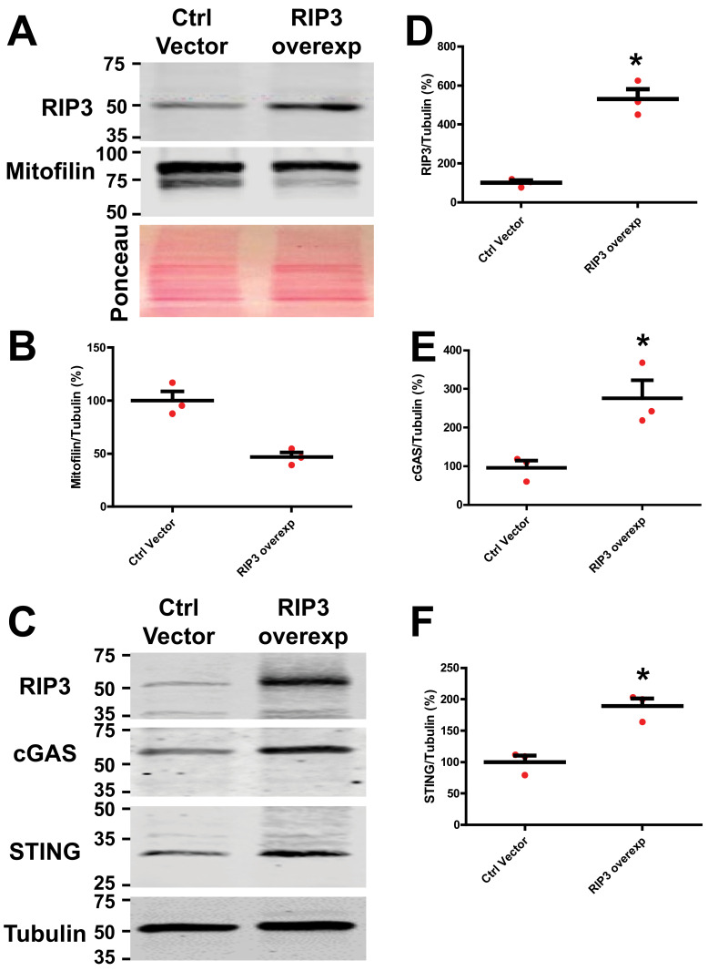
Figure 8.
RIP3 overexpression in HK-2 cells increases the levels of cGAS and STING and reduces Mitofilin levels. (A) Immunoblots and graph (B) showing reductions in Mitofilin levels in HK-2 cells transfected with an RIP3-overexpressed plasmid compared to those transfected with pCMV6 control vector. (C) Immunoblots showing increases in the levels of cGAS and STING levels in HK-2 cells transfected with an RIP3-overexpressed plasmid compared to those transfected with pCMV6 control vector (D–F). Graph showing increases in the levels of RIP 3 (D), cGAS (E), and STING (F) in HK-2 cells transfected with an RIP3-overexpressed plasmid compared to those transfected with pCMV6 control vector. Values are expressed as means ± SEM; * p < 0.05 versus control (Ctrl) vector group (n = 3/group).

