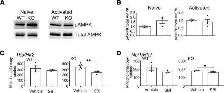
Figure 7. Activation of AMPK maintains mitochondrial mass in Hvcn1-deficient CD8+ T cell activation.
(A and B) Purified naive or Ab-activated (4 days) CD8+ WT and Hvcn1-deficient T cells were lysed and analyzed by Western blotting for the presence of phosphorylated and total AMPK. Quantification (pAMPK/total AMPK) is shown in B (n = 3). (C and D) Total DNA was isolated from Ab-activated (4 days) WT and Hvcn1-deficient CD8+ T cells (n = 3–4) cultured in the presence of the AMPK inhibitor SBI or vehicle alone. Quantitative PCR was used to assess expression of mitochondrial genes 16s in C and Nd1 in D and normalized to the nuclear gene Hk2 to calculate mitochondrial DNA copy number. Data are presented as mean ± SEM (n > 3). Student’s 2-tailed t test; *P < 0.05, **P < 0.01.

