Figure 1.
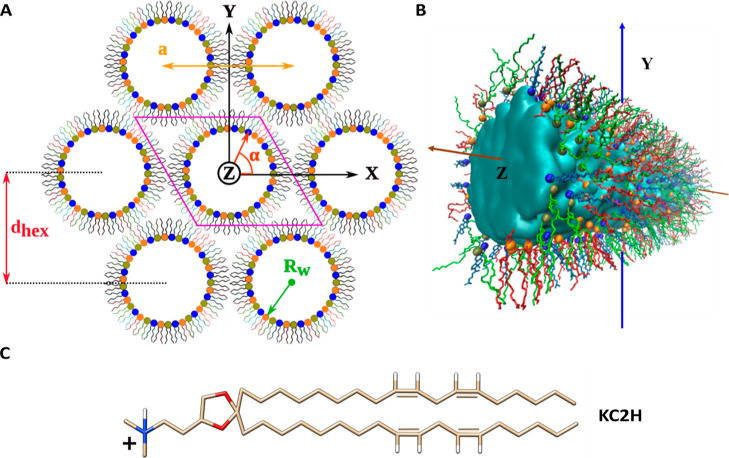
Lipid arrangement in the HII phase (A) HII structure (ternary mixture): lipid tubules filled with water and ions are arranged in a hexagonal geometry. The solvent is not shown for clarity. Each lipid cylinder is perpendicular to the XY plane and parallel to the Z-axis. Lipids are initially distributed radially and randomly around each water core. A quasi-infinite HII lattice can be simulated using a single lipid tubule confined in a triclinic simulation box (shown in pink) and its periodic copies (six are shown). The radius of the water core (Rw), lattice plane distance (dhex), and lattice spacing (a) parameters are illustrated. The X and Y axes point to two of the interaxial and interstitial directions, respectively. (B) Molecular view of the central lipid tubule taken from a simulated DSPS/KC2H/cholesterol (35/35/30) system with 30 nw. The cylinder is rotated ca. 45° around the Y-axis for visualization purposes. The solvent is represented as the cyan surface. DSPS, KC2H, and cholesterol are shown as red, green, and light blue lines. Spheres colored in orange, tan, and dark blue represent the phosphorous atom of DSPS, nitrogen atom of KC2H, and oxygen atom of cholesterol, respectively. Hydrogens are not shown for clarity. VMD52 was used to create the figure. (C) Chemical structure of the KC2H lipid. Oxygen, nitrogen, and hydrogen atoms are colored in red, blue, and white, respectively, whereas the carbon atoms are colored in tan. The two unsaturated bonds in each tail are shown explicitly.

