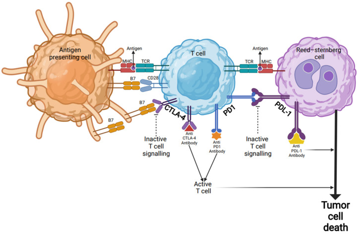
Figure 1.
Checkpoint inhibitor immunotherapy in Hodgkin’s lymphoma. Inactive T cells are activated through their TCR by encountering antigenic peptides presented by the MHC complex on the surfaces of APCs or tumor cells (Reed-Sternberg cells). In addition to TCR-MHC engagement, a co-stimulatory signal via B7 protein is required for target-cell lysis and effector cell responses. B7 protein on activated APCs can pair with either a CD28 on the surface of a T-cell to produce a costimulatory signal to enhance the activity of TCR-MHC signal and T-cell activation, or it can pair with CTLA-4 to produce an inhibitory signal to keep the T cell in the inactive state. Blocking the binding of B7 to CTLA-4 with an anti-CTLA-4 antibody allows T cells to be activated and to kill tumor cells. Other immune checkpoint proteins such as PD-1 on the surfaces of T cells and PD-L1 on tumor cells can also prevent T cells from killing tumor cells. Immune checkpoint blockade via monoclonal antibodies (anti-PD-L1 or anti-PD-1) can lead T cells to kill tumor cells. Abbreviations: MHC: major histocompatibility complex; APC: antigen-presenting cells; TCR: T-cell receptor; CTLA-4: cytotoxic T-lymphocyte associated antigen 4; PD-1: programmed death 1; PD-L1: programmed death-ligand 1.

