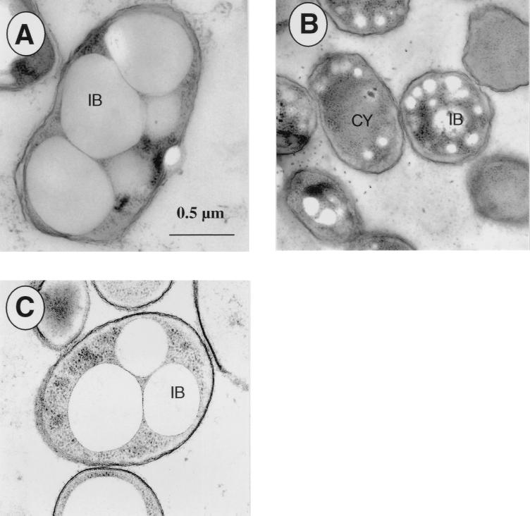
FIG. 2.
Morphology of P. oleovorans GPo1000 and GPo1001. Cells were cultivated in 0.1NE2 minimal medium containing 15 mM octanoate and were harvested in the stationary growth phase. Electron micrographs were obtained as described in Materials and Methods and depict representative cells. (A) P. oleovorans GPo1000. (B) P. oleovorans GPo1001. (C) P. oleovorans GPo1001(pHAD5). CY, cytoplasm; IB, PHA granule (inclusion body). The bar applies to all panels.

