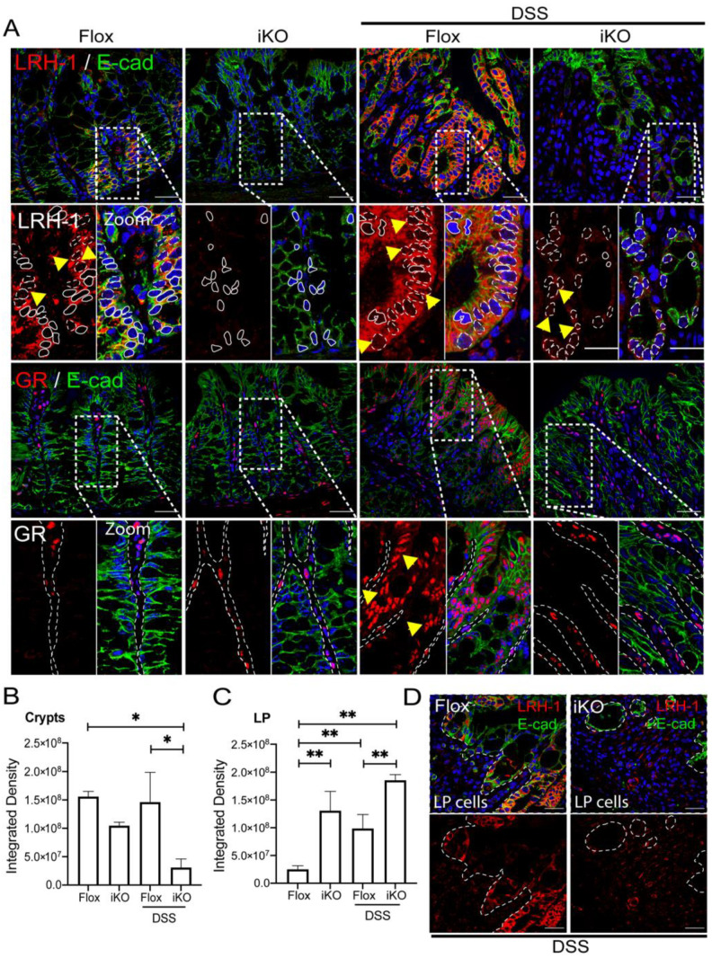
Figure 4.
LRH-1 upregulated in intestinal mucosa lamina propria from DSS-treated GRiKO mice. (A) Representative images of LRH-1 and total GR (red) immunofluorescent staining with co-localization of E-cadherin (green) as epithelial marker in intestinal mucosa from vehicle and DSS-treated GRflox and GRiKO mice. Hoechst was used for nuclear counterstaining. In the LRH-1 zoomed image: dashed line and/or yellow arrows: positive nuclear stain; solid line: negative nuclear stain. In the GR zoomed image, yellow arrows: positive nuclear stain; dashed line: epithelial outline. Objective 60×. Scale bar 30 μm. (vehicle n = 4 GRflox and 3 GRiKO, DSS-treated n = 4 GRflox and 6 GRiKO); (B) Integrated density from immunofluorescence images calculated from epithelial crypts; and (C) LP cells from intestinal mucosa of vehicle and DSS-treated GRflox and GRiKO mice, with (D) a representative image showing LRH-1 stain in LP from DSS-treated groups. Dashed line: epithelial outline. * p < 0.05, ** p < 0.01.

