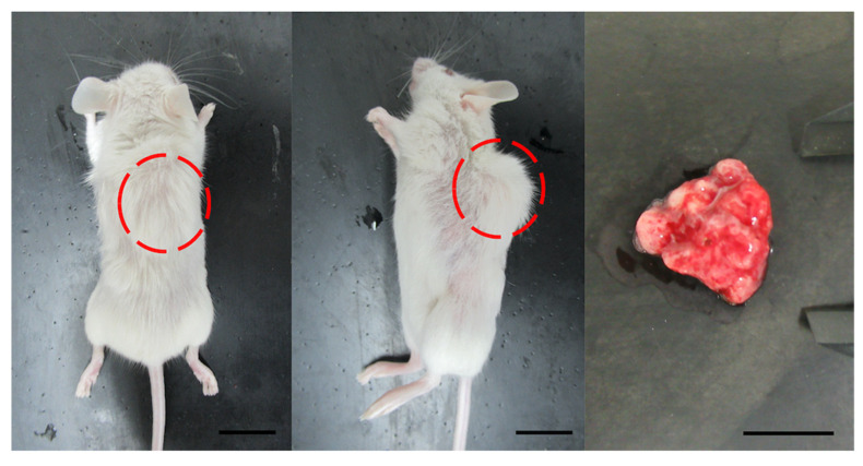
Figure 1.
Development of tumors from a cervical cancer xenograft model (PDX71). Left, whole body image from the back. Solid tumor is evident (red circle). Center, whole-body image from lateral view. The tumor consists of a single nodule (red circle). Right, isolated tumor. Scale bar in all images = 10 mm.

