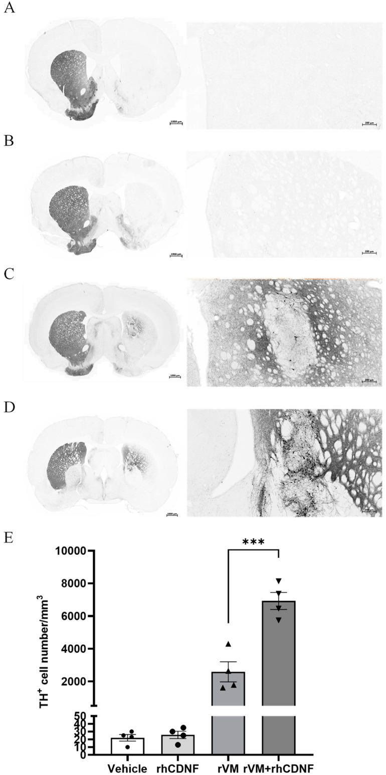
Figure 4.
Photomicrographs of tyrosine hydroxylase immunoreactive (TH−ir) cells in hemiparkinsonian rat striatum at 8 weeks following transplantation. TH-ir cell bodies and fibers in the grafted side (right side of coronal brain sections) of the rVM and rVM + rhCDNF groups. Higher density of TH−ir cells was found in the rVM + rhCDNF group as compared to the rVM group. (A) Vehicle group. (B) rhCDNF alone group. (C) rVM group. (D) rVM + rhCDNF group. (E) Quantification of TH−ir cell density. *** p < 0.001 vs. rVM and rVM + rhCDNF. N= 4–5 rats in each group.

