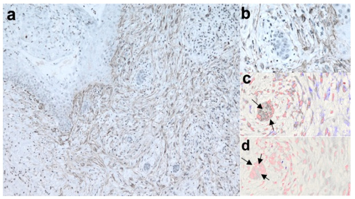
Figure 1.
Interaction between epithelial cells and myofibroblasts during connective wall invasion in NBCCS-OKC. (a), αSMA staining depicts myofibroblasts surrounding invading epithelial droplets (LSAB-HRP, ×10; nuclear counterstaining with hematoxylin); (b), further magnification of a (LSAB-HRP, ×40); invading droplets have been further examined on serial sections by digital pathology showing high expression of MMP-7; (c), digital pathology, ×40, MMP-7 in violet and nuclear staining in red pseudocolors); (d), digital pathology ×40, MMP-9 in violet and nuclear staining in red pseudocolors), whereas MMP-9 showed a low level of expression [arrows indicated area of epithelial nuclei].

