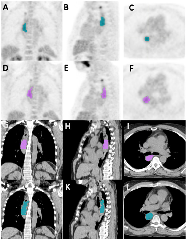
Figure 2.
Coronal (A,D), sagittal (B,E), and axial (C,F) PET images, and coronal (G,J), sagittal (H,K), and axial (I,F) CT images showing segmentation of primary tumor by reader 1 (A–C,G–I) and reader 2 (D–F,J–L), in a 64-year-old male patient with esophageal squamous cell carcinoma; the Jaccard index and a dice similarity coefficient for inter-reader agreement on the segmented volumes were 0.657 and 0.793, respectively.

