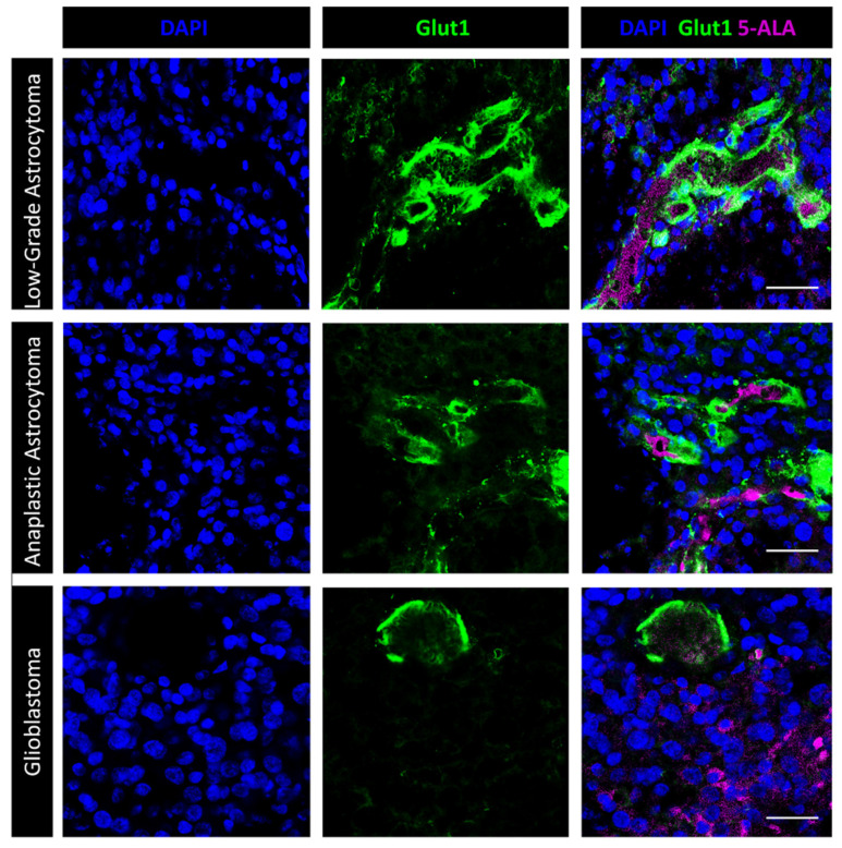
Figure 5.
Fluorescence microscopy for 5-ALA and immunofluorescence with anti-Glut1. In low grade glioma (upper panels; Case #2 on Table 1), the dotted 5-ALA fluorescent signal is mostly confined to intravascular cells. A few fluorescent dots can be seen in perivascular cells. Glut1 is highly expressed by the endothelial cells. Scale bar, 40 μm. In anaplastic glioma (middle panel; Case #4 on Table 1), the Glut1 staining lacks in many regions of the tumor vessels, suggesting BBB disruption. Mosaic and/or perivascular tumor cells show intense 5-ALA fluorescence. Scale bar, 40 μm. Glioblastoma tumors (lower panels; Case #14 on Table 1) show several 5-ALA fluorescent cells even at distance from vessels with discontinuous Glut1 staining. Scale bar, 40 μm. Objective lens, Olympus PlanApo N 60×/1.42 Oil.

