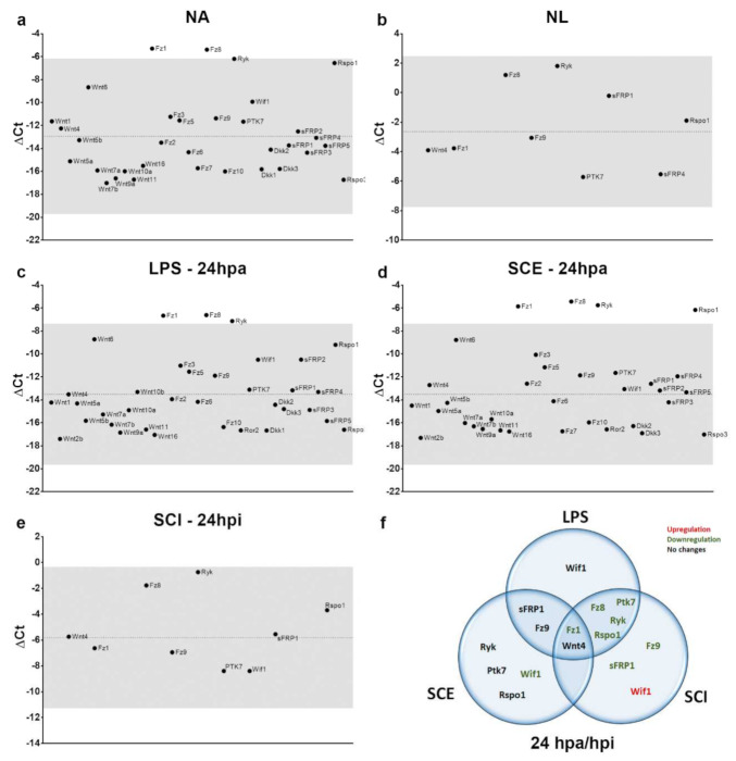
Figure 6.
Schematic overview of harmful stimulus effect on microglial cells both in vitro and ex vivo for the three experimental conditions that were evaluated at 24 h post-treatment/injury. Graphic representation of ΔCt values for the different genes that were analyzed in the different evaluated conditions, where the dashed line indicates ΔCt mean value in each case and gray shading identifies an area corresponding to the mean value ± 2 times the standard deviation to highlight those genes with higher/lower expression levels for each experimental condition. (a) Wnt-related genes that were detected in non-activated microglial cells in vitro, (b) Wnt-related genes that were detected in microglial cells isolated from non-lesioned spinal cord, (c) Wnt-related genes that were detected in LPS-activated microglial cells in vitro at 24 hpa, (d) Wnt-related genes that were detected in SCE-activated microglial cells in vitro at 24 hpa, (e) Wnt-related genes that were detected in microglial cells that were isolated from injured spinal cord at 24 hpi, (f) Venn diagram shows individual and shared gene alterations that were observed for the three experimental conditions that were evaluated at 24 h post-activation/injury. NA: non-activated; NL: non-lesioned; LPS: lipopolysaccharide; SCE: spinal cord extract; SCI: spinal cord injury; hpa: hours post-activation; dpa: days post-activation; hpi: hours post-injury.

