Figure 8.
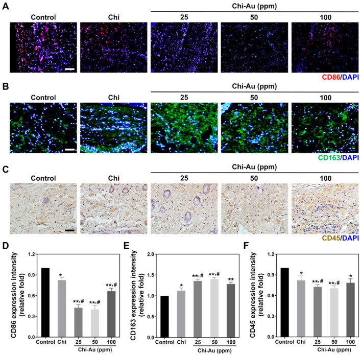
Immunohistochemical (IHC) staining for the investigation of anti-inflammatory capacities after subcutaneous implantation for 4 weeks. (A,B) The expressions of CD86 (M1 polarization, red color) and CD163 (M2 polarization, green color) are shown. (C) The expression of CD45, which indicates leukocyte filtration, was observed using DAB staining. (D,E) The expressions of CD86 and CD163 were semi-quantified. Chi combined with 50 ppm of Au stimulated the lowest CD86 expression. In contrast, the expression of CD163 was remarkably greater with Chi–Au 50 ppm treatment. (F) The CD45 expression was quantified. Based on the result, Chi–Au 50 ppm induced the lowest CD45 expression. The results elucidate that Chi with 50 ppm of Au exhibited better biocompatibility when compared with the other tested nanocomposites. Scale bar was 100 μm. Results are presented as mean ± SD (n = 3). * p < 0.05, ** p < 0.01: compared with the control (TCPS); # p < 0.05: compared with the Chi group.

