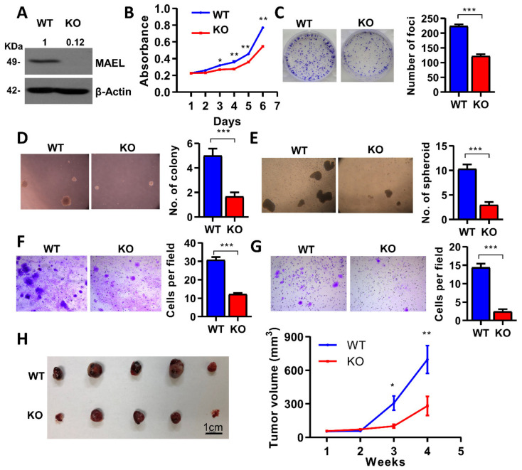
Figure 2.
Deletion of MAEL-impaired aggressive phenotype in HCC. (A) Western blot validation of MAEL knockout in PLC8024 cells. β-Actin served as an internal control. Full Western Blot can be found in Figure S6. (B) Cell proliferation rate determined by XTT assay. Representative images and quantitation of (C) foci formation, (D) colony formation, and (E) spheroids in soft agar in MAEL wildtype and knockout PLC8024 cells. Representative images and quantitation of (F) migrated and (G) invaded cells in MAEL wildtype and knockout PLC8024 cells. (H) Representative images and tumor volumes of xenograft tumors derived from mice subcutaneously inoculated with wildtype or knockout cells (n = 5 per group). WT, wildtype; KO: MAEL knockout. Scale bar stands for 1 cm. The values indicate the mean ± SD of three independent experiments. * p < 0.05; ** p < 0.01; *** p < 0.001.

