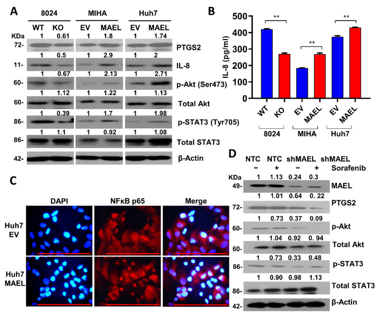
Figure 7.
MAEL activates the IL8/Akt//NFκB/STAT3 signaling pathway. (A) Immunoblots of PTGS2, IL-8, STAT3, p-STAT3, AKT, and p-Akt in cells with or without modulated MAEL expression. Full Western Blot can be found in Figure S9. (B) Quantitative analysis of IL-8 concentration determined by ELISA in MAEL expression-modulated cells. (C) Representative images of immunofluorescence of Huh7 cells transfected with MAEL or vector cells. Cells were stained with NF-κB (P65) antibody (red). Nuclei were labeled by DAPI (blue). Scale bar stands for 50 µm. (D) Immunoblots of pAkt, pSTAT3, and PTGS2 in PLC8024 cells treated with sorafenib with or without MAEL silencing. Data represent the mean ± SD of three independent experiments. ** p < 0.01. Full Western Blot can be found in Figure S10.

