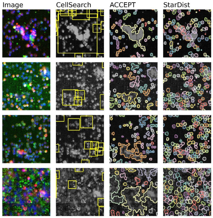
Figure 1.
Four example images with the accompanying segmentations as performed by CellSearch, ACCEPT, and StarDist. The first column shows a false-color image in which the nuclear staining is colored blue (DAPI), the cytokeratin staining green (CK-PE), and CD45 staining red (CD45-APC). The second column shows the same image in black and white with the yellow rectangular CellSearch segmentation areas. In the third and fourth columns, the ACCEPT and StarDist segmentations are depicted as a separate color for each segmented event. StarDist clearly performs best in segmenting all single events separately, especially in areas containing a high cell density.

