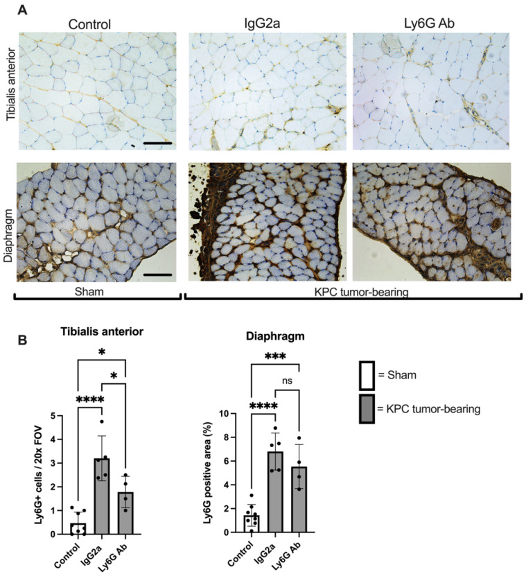
Figure 4.
Increased presence of Ly6G+ cells in limb muscles of KPC mice is blunted with anti-Ly6G Ab treatment. (A) Representative cross-sectional images of tibialis anterior and diaphragm muscles from Sham (Control; n = 8) and KPC mice treated with either isotype control antibody (n = 5) or anti-Ly6G antibody (n = 4) were stained for the Ly6G antigen. Images were analyzed and are quantified in panel (B). Four randomly selected 20× images were analyzed for each mouse of each group. Open bars = Sham (Control). Grey bars = KPC. * p < 0.05, *** p < 0.001, **** p < 0.001, ns = not significant. Scale bars = 100 µm.

