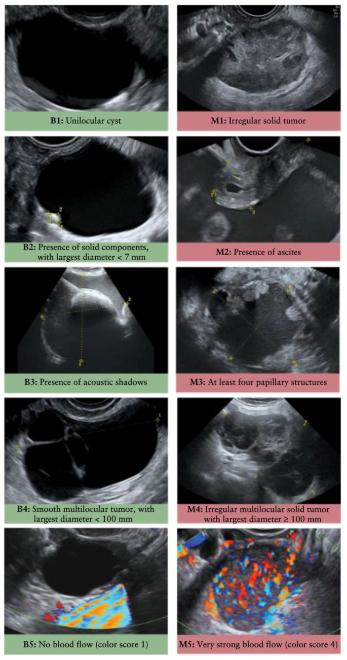
Figure 2.
Representative sonographic images of benign (B1–B5) and malignant (M1–M5) ovarian masses. Features in each panel are reviewed based on the IOTA simple rules to evaluate malignancy of ovarian lesions. In general, the presence of irregular shaped bodies, papillary projections, and/or internal blood flow are predictive of malignancy. Images provided from Kaijser et al. [41]. Used with permission.

