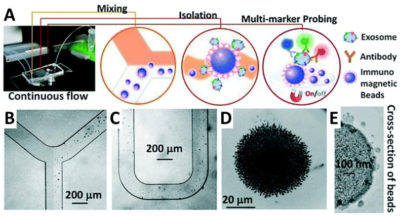
Figure 9.
Workflow of the ExoSearch chip developed by Zhao et al. [257]. (A) Patient plasma (orange) is mixed with immunomagnetic beads that bind exosomes within the sample. Beads carrying exosomes are then isolated via a magnetized field in which the number of beads isolated was in direct comparison to the sample input and could be quantified. A mixture of fluorescently labeled antibodies is then applied to the isolated beads for multi-color fluorescence imaging. (B,C) Bright-field images of the immunomagnetic beads in the microfluidic compartments. (D) Aggregated exosome-bound immunomagnetic beads after magnetic separation (E) Transmission electron micrograph depicting the cross-section of an exosome-bound immunomagnetic bead. Reprinted under the Creative Commons CC BY-NC3.0 license.

