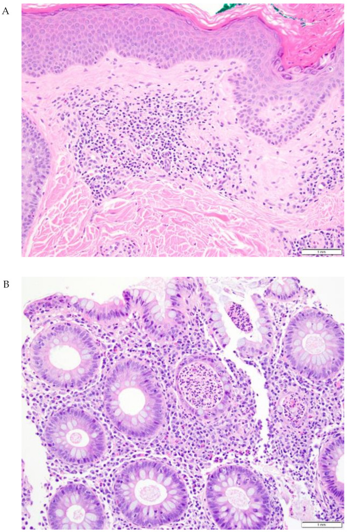
Figure 3.
Representative skin and gut biopsies (20×) from a patient with both ircAE and irColitis. (A) Lichenoid dermatitis with eosinophils and focal acantholytic dermatosis. (B) Active colitis with crypt abscesses, scattered intraepithelial lymphocytes, and increased inflammatory cells in lamina propria.

