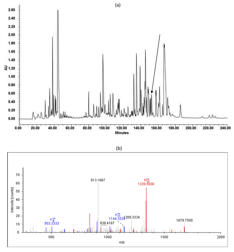
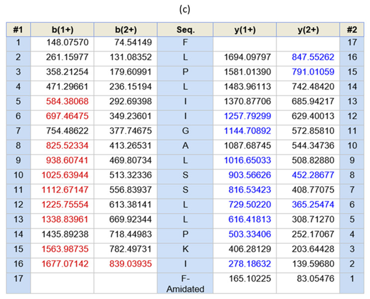
Figure 2.
(a) The RP-HPLC chromatogram of the skin secretion of Pelophylax kl. esculentus. The peak of temporin-PKE is indicated by an arrow. (b) The annotated LCQ tandem mass (MS/MS) fragmentation spectrum of temporin-PKE. Prominent ions in are labeled with the observed m/z value and ion series designation. b and y denote the N- and C-terminal fragments of the peptide produced by breakage at the peptide bond in LCQ, respectively. The number represents the residue number from either the N- or C- terminus. (c) Electrospray ion-trap MS/MS fragmentation data derived from fragment ions in panel (b) corresponding in molecular mass to temporin-PKE. Predicted singly- and doubly-charged ions are coloured black. The actual b-ions and y-ions detected by MS/MS fragmentation are indicated in red and blue typeface, respectively.


