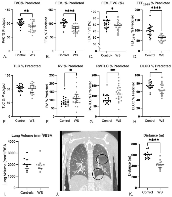
Figure 1.
Pulmonary evaluation reveals reduced lung function and air trapping in cases compared to controls. Spirometry and lung volume measurements in WS cases and healthy controls: (A) FVC, (B) FEV1, (C) FEV1/FVC, (D) FEF25–75, (E) TLC, (F) RV, (G) RV/TLC, and (H) DLCO are shown (all are presented as percentage predicted values except FEV1/FVC [%]). All data are presented as median and interquartile range. 21/22 cases completed RV, RV/TLC, and DLCO testing. Statistics are reported as Mann–Whitney tests. p values reported are * p < 0.05, ** p < 0.01, and **** p < 0.0001. CT analyses show adults with WS have normal lung volumes: (I) Lung volumes assessed via CT and normalized to BSA, but a subset show evidence of air trapping: (J) Representative image of focal lucency in the base of the left lung and an additional heterogeneous area in the left mid-lung in a patient with WS. Statistics are reported as Mann–Whitney test. At the same time, (K) individuals with WS have significantly shorter 6 min walk distances compared to controls. Statistics are reported as a Mann–Whitney test. For all tests: p values reported are * p < 0.05, ** p < 0.01, and **** p < 0.0001. Abbreviations: FVC: forced vital capacity, FEV1: forced expiratory volume in 1 s, FEF25–75: Forced expiratory flow between 25 and 75%, TLC: total lung capacity, RV: residual volume, DLCO: diffusion capacity for carbon monoxide, BSA: body surface area.

