Figure 1.
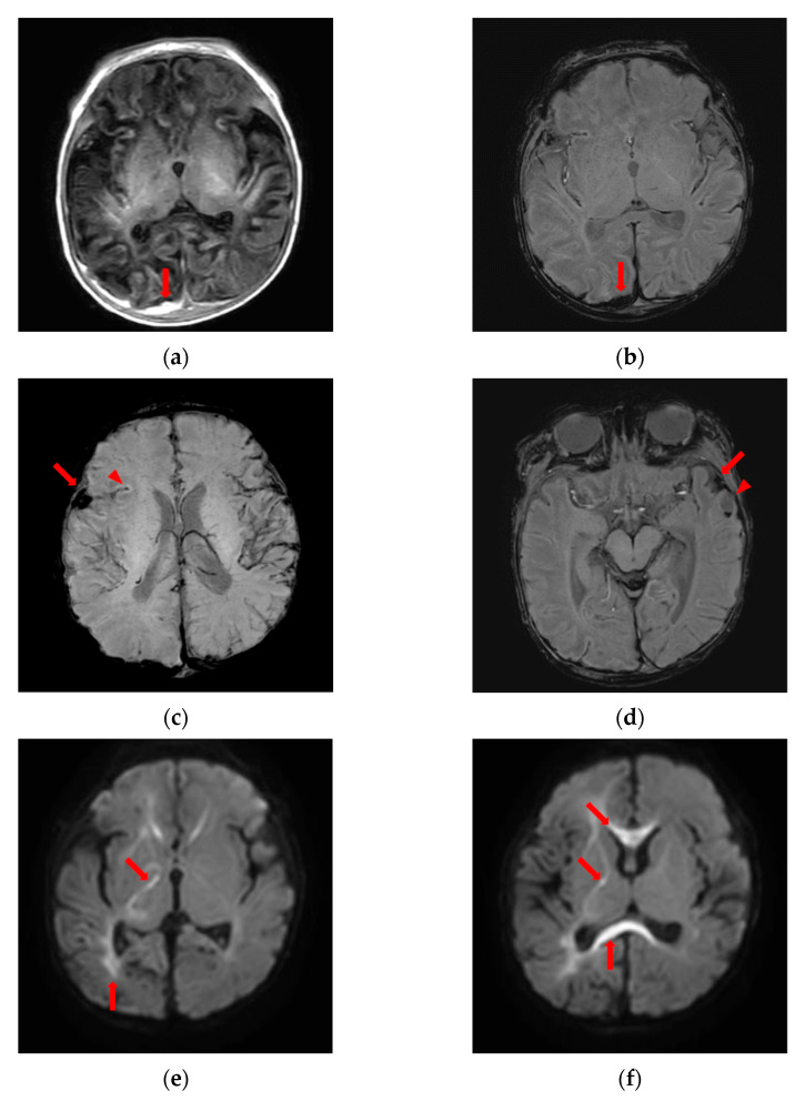
Brain magnetic resonance imaging suggesting abusive head trauma. (a) High signal intensity on T1-weighted image and (b) low signal intensity on susceptibility-weighted image, subdural fluid collection along the posterior aspect of the falx cerebri and occipital convexity implying subacute subdural hemorrhage (arrows); (c) low signal intensity in the right sylvian cistern (arrow), right sylvian fissure (arrowhead) and (d) left sylvian cistern (arrow) implying subarachnoid hemorrhage on susceptibility-weighted image; (d) low signal intensity with fluid-fluid level (arrowhead) along the anterior side of left temporal lobe implying acute hemorrhage on susceptibility-weighted image; (e,f) diffusion restriction in periventricular white matter, corpus callosum, right thalamus, and internal and external capsules on diffusion-weighted image (arrows).

