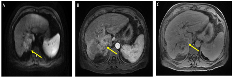
Figure 1.
Magnetic resonance imaging of the abdomen. Limited by motion. Cirrhosis and splenomegaly were noted. Infiltrative segment 7 mass measures 6.5 × 4 cm. (A) Diffusion-weighted imaging (DWI) shows diffusion restriction of mass; (B) T1 with contrast shows vague arterial phase enhancement; (C) T1 post-contrast delayed phase imaging shows washout (captured at the diagnosis visit, and before the atezolizumab plus bevacizumab started).

