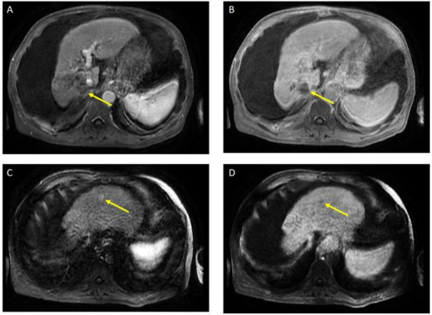
Figure 3.
Magnetic resonance imaging of the abdomen. Post-treatment atrophy of the right hepatic lobe, cirrhosis, splenomegaly, and ascites were noted. T1 post-contrast early (A) and delayed (B) demonstrate phase unchanged Seg 7 non-enhancing 4.5 cm cavity (LR TR nonviable); T1 with contrast early (C) and delayed (D) show new 8 mm lesion in segment 2 with early enhancement and late washout (LR 4) (captured at the follow-up visit, and 6 months after the atezolizumab plus bevacizumab started).

