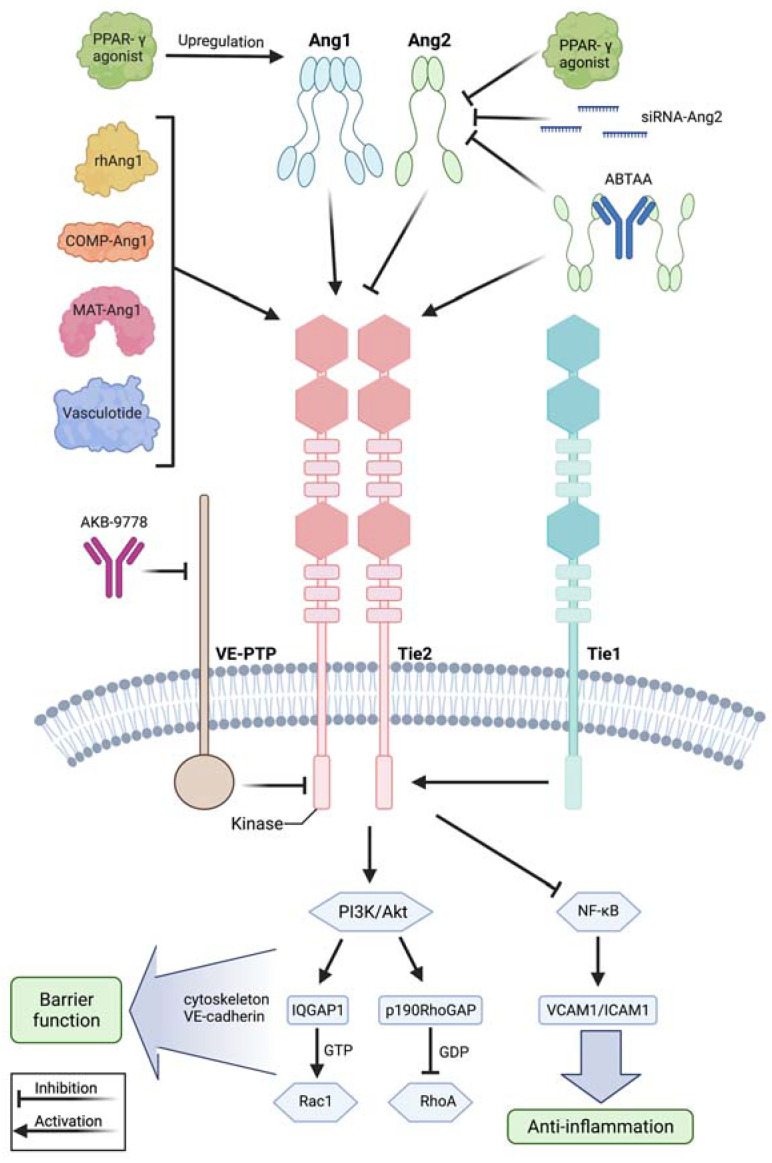
Figure 2.
Modulation of the angiopoietin-Tie2 signaling axis during systemic inflammation. Recombinant human angiopoietin 1 (rhAng1), cartilage oligomeric matrix protein (COMP)-angiopoietin 1 (COMP-Ang1), matrilin-1-angiopoietin-1 (MAT-Ang1) and vasculotide are Tie2 agonists with similar action to angiopoietin 1, while peroxisome proliferator–activated receptor-γ (PPAR-γ) agonists upregulate angiopoietin 1 bioavailability. Small interfering RNA (siRNA) against angiopoietin 2 and PPAR-γ agonists reduce angiopoietin 2 expression. AKB-9778 is an antibody directed against vascular endothelial protein tyrosine phosphatase (VE-PTP) and thus indirectly activates Tie2. ABTAA is a novel ANG2-binding and Tie2-activating antibody combining the features of angiopoietin 2 inhibition and Tie2 activation. Tie1 further modulates response at the Tie2 receptor as endothelial cells shed the Tie1 ectodomain leading to Ang2 binding, resulting in Tie2 antagonism and reducing the agonistic activity of Ang1 during inflammation. Mechanistically, these treatment strategies lead to Tie2 receptor agonism, resulting in enhanced vascular barrier function by the PI3K/Akt signaling cascade and anti-inflammation by suppression of transcription factor NF-κB and, thus, of intercellular adhesion molecule (I-CAM) and vascular cell adhesion molecule (V-CAM). Downstream of phosphatidylinositol-3-kinase/protein kinase B (PI3/Akt) activation, there is the GTPase-activating protein 1 (IQGAP1), which activates Rac1 by stabilizing it in its active GTP-bound form, whereas Rho GTPase-activating protein p190RhoGAP converts RhoA into its inactive state. These steps promote an increase in cortical actin that strengthens the cytoskeleton and thus the vascular barrier function, whereas Tie2 activation additionally results in inhibition of Src kinase preventing the phosphorylation and internalization of vascular endothelial-cadherin (VE-cadherin). This figure was created with BioRender.com and adapted from Pariksh SM et al., J Am Soc Nephr 2017 and Wettschureck et al., Physiol Rev 2019 [36,74].

