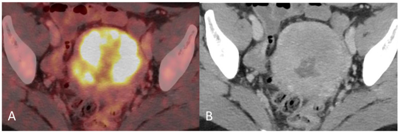
Figure 2.
(A) Baseline axial PET/CT fused image. The metabolic activity is inhomogeneous, with areas of high to low metabolic activity. (B) Contrast-enhanced axial CT shows an enlarged uterus with endometrial thickening and polypoid tumour masses protruding into the uterine cavity. The tumor appears hypodense to the myometrium with signs of myometrial invasion. There was no involvement of the ovaries, fallopian tubes or cervix on either the PET or CT.

