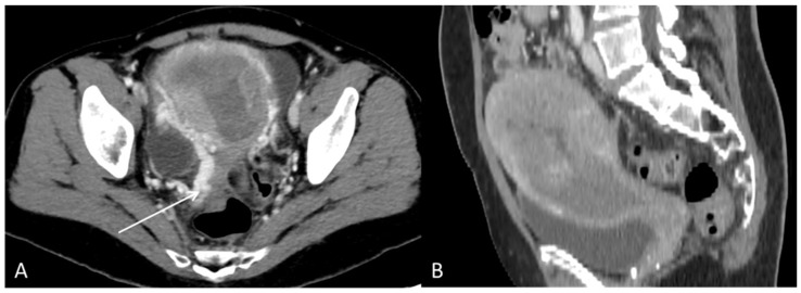
Figure 7.
The CT with contrast enhancement performed after approximately two months of sorafenib treatment showed a morphological progression of the primary tumour in the uterus. During this time, the patient had also received radiation therapy (3Gyx10) of the uterus as palliative treatment due to heavy vaginal bleeding. (A) Axial CT and (B) sagittal CT showed tumour masses in the uterine cavity with patchy hypodensities consistent with necrosis. In the tumour periphery, contrast-enhanced varicose veins were visible, consistent with pelvic congestion syndrome ((A), arrow).

