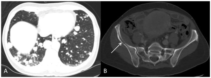
Figure 8.
The contrast-enhanced CT, after approximately two months of sorafenib treatment, showed progression with multiple pulmonary metastases (A) and an osteolytic metastasis with corticalis destruction in the right pelvic bone ((B), arrow). S-AFP was 412,200 kIU/L. Sorafenib was discontinued due to progression. The patient passed away shortly after.

