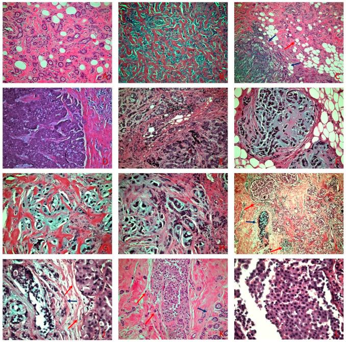
Figure 1.
(A) MGA with small round uniform tubules lined with a single layer of cuboidal epithelial cells with occasional eosinophilic luminal secretion (H&E × 100). (B) AMGA displays areas of greater architectural complexity compared to MGA (H&E × 100). (C) MMPC (blue arrows) adjacent to MGA (red arrow) (H&E × 100) (D) Areas of solid invasive carcinoma adjacent to areas consisting of small nests with an abrupt transition to chondromyxoid matrix (H&E × 100). (E–H) Small nests, cords, single cells, tubular-like structures, and ring-like structures, embedded in a chondromyxoid matrix (H&E × 200). (I) On low power examination, PLCIS (red arrows) is adjacent to MMPC (blue arrow) (H&E × 40). (J) PLCIS (red arrows) shows cells with moderate to severe atypia, eosinophilic granular cytoplasm, and central to eccentric hyperchromatic nuclei adjacent to MMPC (blue arrow) (H&E × 200) (K) PLCIS (red arrows) adjacent to MGA (blue arrow) (H&E × 100). (L) On high power examination, lack of cellular cohesion is evident in the PLCIS (H&E × 400).

