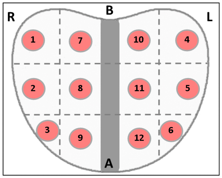
Figure 1.
Prostate divided into twelve target zones, resulting a total of twelve biopsy fragments at approximatively 1 cm distance between each other. Every target zone and tissue fragment corresponds to a region evaluated with SWE measurements before the biopsies were taken (R = right, L = left, B = base and A = apex).

