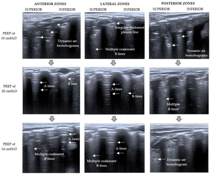
Figure 3.
Left LUS examination during RMs. The point where the LUS probe is applied is indicated at the top of the figure; on the left, the PEEP level at that moment is specified. Baseline LUS (PEEP of 10 cmH2O) showed a C pattern (severe loss of aeration with dynamic air bronchograms). With a PEEP of 30 cmH2O, the pattern changed from C to B1 (moderate loss of lung aeration with multiple well-defined B-lines and some A-lines). After RMs (PEEP of 14 cmH2O), LUS showed a severe loss of lung aeration with multiple coalescent B-lines but some A-lines in the anterior and lateral zones (B2 pattern).

