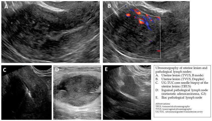
Figure A5.
(A,B) Transvaginal ultrasonogram of the uterine lesion (atypical) in the myometrium in the uterine doom, B mode and Doppler. (C) Transrectal ultrasonogram of the UG-TUC core needle biopsy of the myometrial lesion. (D) Ultrasonogram (linear probe) of the pathological inguinal lymph node. (E) Transvaginal ultrasonogram of the pathological iliac lymph node. Histology: leiomyoma (uterine lesion), normal endometrium (from curettage), adenocarcinoma G3 in the inguinal lymph node (biopsy).

