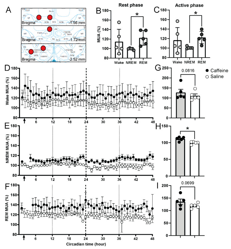
Figure 5.
Electrical activity in lateral hypothalamus under caffeine and saline treatment over 48 h. The location of the electrodes in PLH is shown in (A). PLH activity under subjective day (B) and subjective night (C) under baseline condition (saline-treated). CT0-CT12 was considered as the subjective day, CT12-CT24 was considered as the subjective night. Asterisks indicate significant neuronal activity differences between NREM and REM states (* p < 0.05, paired t-test). (D–F) Time course of neuronal activity of PLH in waking, NREM, and REM sleep (2 h interval) for caffeine (black, n = 5) and saline (white, n = 5) administration in 48 h. The first 24 h was considered as treatment day; the second 24 h was considered as the recovery day. Arrow indicates the injection time (CT1-2). (G–I) Average value of 47 h of neuronal activity of PLH in waking, NREM, and REM sleep for caffeine (black, n = 5) and saline (n = 5). Asterisk indicates significant differences between caffeine and saline (* p = 0.0251, paired t-test). Data are shown as mean ± SEM.

