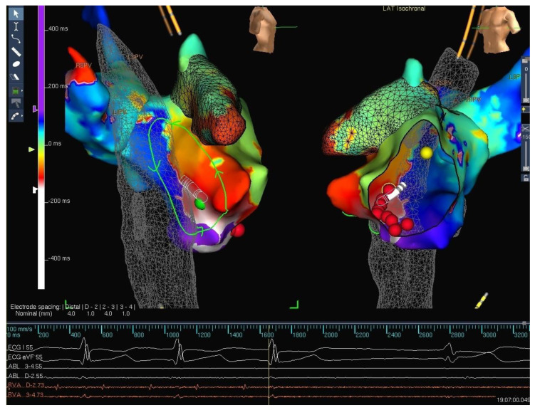
Figure 4.
Three-dimensional electroanatomical mapping of a patient with single ventricular physiology post Fontan palliation with intra atrial re-entrant tachycardia evident from the activation mapping. The non-colored part of the mapping shell is representative of the lateral tunnel. The non-standard projection in this image is to show the important part of the circuit and geometry, again indicating the ability to view the geometry from any projection as deemed necessary.

