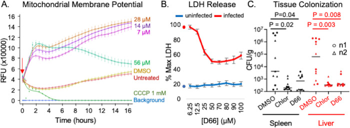
Fig 5. D66 is well tolerated by eukaryotic cells and has antimicrobial activity in mice.
A) RAW 264.7 cells were incubated with the mitochondrial membrane potential indicator TMRM, treated (red arrow) with DMSO (0.5%), CCCP, or dilutions of D66, and imaged over time. Averages and SEM of three biological replicates with technical triplicates, normalized to time 0. B) RAW 264.7 cells that were uninfected or infected with S. Typhimurium SL1344 for 2 hours were treated with DMSO or D66 and monitored for LDH release after 16 hours. Averages and SEM of three biological replicates with technical duplicates, normalized to the maximum amount of LDH release (lysed cells; % Max LDH). Symbols on the Y-axis show the percentage of LDH released by DMSO- treated cells. C) C57Bl/6 mice were intraperitoneally inoculated with S. Typhimurium. At 10 minutes and 24 hours after infection, mice were dosed with 50 mg/kg of chloramphenicol or D66 by intraperitoneal injection. Mice were euthanized 48 hours after infection. The spleen and liver were homogenized and plated for enumeration of CFU. Significance was determined by Mann-Whitney.

